
From Wikipedia, the free encyclopedia
Content deleted Content added
|
|
|||
| Line 28: | Line 28: | ||
|
==Diagnosis== |
==Diagnosis== |
||
|
[[Computerized tomography]] is the ideal for typifying facet joint arthrosis; evidence suggests that [[magnetic resonance imaging]] is not as sensitive in identifying bony changes.<ref>{{Cite book|title=iSpine: Evidence-Based Interventional Spine Care|others=Michael J. DePalma, MD(editor)|publisher=Demos Medical Publishing|date=2011|isbn=9781935281931}}</ref> |
[[Computerized tomography]] is the ideal for typifying facet joint arthrosis; evidence suggests that [[magnetic resonance imaging]] is not as sensitive in identifying bony changes.<ref>{{Cite book|title=iSpine: Evidence-Based Interventional Spine Care|others=Michael J. DePalma, MD(editor)|publisher=Demos Medical Publishing|date=2011|isbn=9781935281931}}</ref> |
||
|
==Management== |
|||
|
{{Empty section|date=December 2017}} |
|||
|
==See also== |
==See also== |
||
Latest revision as of 20:58, 19 November 2025
Medical condition
| Facet joint arthrosis | |
|---|---|
 |
|
| Facet joints | |
| Specialty | Orthopedic |
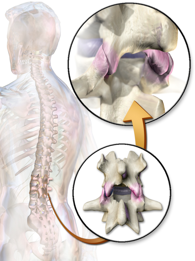
Facet joint arthrosis is an intervertebral disc disorder. The facet joints or zygapophyseal joints are synovial cartilage covered joints that limit the movement of the spine and preserve segmental stability. In the event of hypertrophy of the vertebrae painful arthrosis can occur.[1] The “lumbar facet arthrosis syndrome” was described in a 1987 article by S. M. Eisenstein and C. R. Parry of Witwatersrand University.[2]
Computerized tomography is the ideal for typifying facet joint arthrosis; evidence suggests that magnetic resonance imaging is not as sensitive in identifying bony changes.[3]